Journal of Surgery and Surgical Research
Rare Case Reports on Ectopic Liver Tissue: Incidence, Surgical Management, and Outcomes
Mohamed Dheker Touati1,2*, Ahmed Bouzid1,2, Wassim Romdhane1,2, Fahd Khefacha1,2, Med Raouf Ben Othmane1,2, Wassim Dziri1,2, Anis Belhadj1,2, Ahmed Saidani1,2 and Faouzi Chebbi1,2
1General Surgery Department, Mahmoud El Matri Hospital, V59M+628, Ariana, Tunisia
2Faculty of Medicine of Tunis, University of Tunis El Manar, R534+F9H, Street of the Faculty of Medicine, Tunis, Tunisia
Cite this as
Touati MD, Bouzid A, Romdhane W, Khefacha F, Ben Othmane MR, Dziri W, et al. Rare Case Reports on Ectopic Liver Tissue: Incidence, Surgical Management, and Outcomes. J Surg Surgical Res. 2024;10(2):039-041. Available from: 10.17352/2455-2968.000165Copyright
© 2024 Touati MD, et al. This is an open-access article distributed under the terms of the Creative Commons Attribution License, which permits unrestricted use, distribution, and reproduction in any medium, provided the original author and source are credited.Introduction and importance: Ectopic liver tissue is a rare finding, often discovered incidentally during procedures like cholecystectomy. Understanding its clinical implications, including potential malignancy and complications, is crucial for effective management and improving patient outcomes.
Case presentation: A 45-year-old female presented with six months of biliary colic, worsened by fatty meals. Preoperative ultrasound revealed a gallbladder with microcalculi. During elective laparoscopic cholecystectomy, a brownish tissue fragment resembling hepatic parenchyma was found on the gallbladder fundus and removed. Histopathology confirmed ectopic liver tissue with mild steatosis and no malignancy.
Clinical discussion: Ectopic liver tissue, with a prevalence of 0.47%, typically attaches to the gallbladder but can also be found in other abdominal and thoracic locations. It may be linked to congenital malformations and has a risk of degeneration into malignancy due to its incomplete vascular and ductal system. Diagnosis is usually incidental during surgery, and en-bloc removal is advised to prevent complications and potential neoplastic transformation. Identifying its vascular supply before dissection is crucial to avoid severe bleeding.
Conclusion: Ectopic liver tissue, despite its rarity, requires careful management due to its potential for malignancy and complications. Timely detection and en-bloc removal are vital to prevent adverse outcomes and ensure optimal patient care.
Introduction
Ectopic liver tissue is a rare anatomical finding, with a reported prevalence of approximately 0.47% [1]. It is most commonly associated with the gallbladder but can also be observed in various other intra-abdominal and intra-thoracic sites. Ectopic liver tissue may occasionally present during routine procedures, such as cholecystectomy, and is generally discovered incidentally. Given the risk of malignancy and associated complications, such as hemorrhage or rupture, prompt identification and en-bloc removal are essential. This article aims to review the incidence, clinical implications, and management strategies for ectopic liver tissue, emphasizing the importance of recognizing and addressing this anomaly to ensure optimal patient outcomes.
This work has been reported in line with the SCARE 2023 criteria [2].
Case presentation
A 45-year-old female, with no significant medical or surgical history, presented with a six-month history of biliary colic, exacerbated by fatty meals and relieved by antispasmodics. The interview did not reveal any recent jaundice or other associated functional symptoms. The physical examination showed normal conjunctivae, and the abdominal examination was unremarkable. Laboratory tests, including liver function tests, were within normal limits. Preoperative abdominal ultrasound revealed a non-distended gallbladder with a thin wall containing multiple microcalculi. The bile ducts were normal in size, and the liver appeared normal in morphology and size.
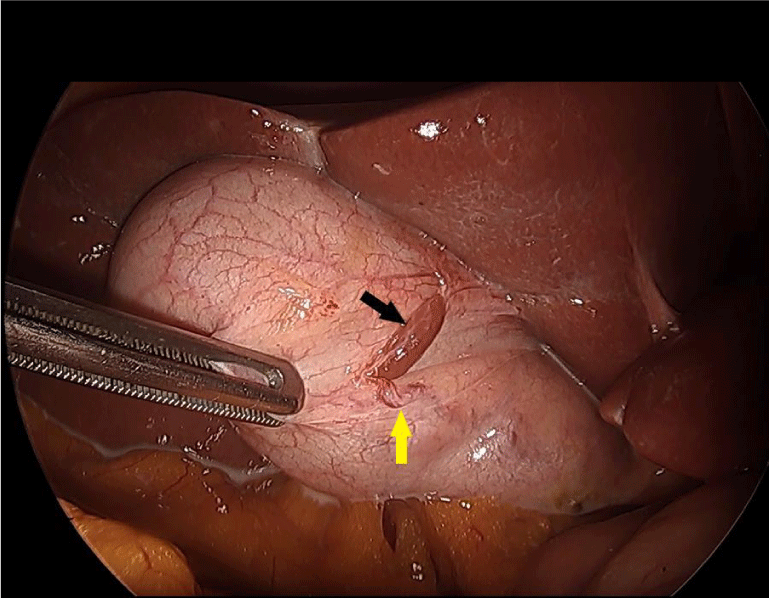
The patient was scheduled for an elective laparoscopic cholecystectomy. Intraoperative exploration showed a non-distended gallbladder with a thin wall. A centimeter-sized brownish tissue fragment, resembling hepatic parenchyma, was identified on the anterior wall of the gallbladder fundus. Its distinct color facilitated identification. The fragment measured 1 cm (Figure 1) and had an independent vascular pedicle originating from the cystic artery, with no connection to the main biliary system or cystic duct. The vascular pedicle was isolated and clipped separately, allowing for a retrograde cholecystectomy with the removal of the tissue fragment. The main difficulty during the procedure was to resect the ectopic liver tissue en bloc with the gallbladder and carefully dissect and control its vascular pedicle.
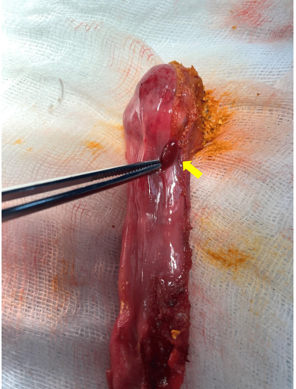
Upon opening, the gallbladder contained multiple microcalculi. The excised specimen was sent for histopathological analysis (Figure 2). The analysis confirmed the presence of ectopic liver tissue adhering to the gallbladder serosa, with sinusoidal congestion, mild steatosis, and focal hemosiderin deposits, without evidence of malignant degeneration. The postoperative course was uneventful, and the patient was discharged on postoperative day 1 with a favorable outcome.
Follow-up examinations were conducted at regular intervals over a year, and no complications or abnormalities were detected. The patient remained asymptomatic, with normal laboratory and imaging findings throughout the entire follow-up period, indicating a complete and sustained recovery.
Discussion
The incidence of ectopic liver tissue is particularly low, with a reported prevalence of 0.47% [1]. Several theories have been proposed to explain the presence of this ectopic tissue [3]. However, it is widely accepted that it forms during the fourth week of embryonic development in utero, due to the displacement of a portion of the cranial part of the hepatic diverticulum from the liver bud to other locations [4]. The ectopic liver is generally attached to the serosa of the gallbladder or embedded within its wall, but it can also be found in the lumen of the gallbladder [5].
Ectopic liver tissue is sometimes associated with other congenital malformations, such as biliary atresia, agenesis of the caudate lobe, omphalocele, biliary cysts, or cardiac anomalies [5]. Although the gallbladder is the most common site for this tissue, it can also be located in the intra-abdominal and intra-thoracic cavities[6]. Cases have been observed in the spleen, umbilicus, and vena cava, as well as in the heart and lungs [6,7].
Ectopic liver tissue should be removed due to its potential to undergo neoplastic transformation, regardless of the condition of the main liver [8]. This tissue is considered more likely to become malignant because it lacks a complete vascular or ductal system, which can lead to functional impairments. Altered liver function can result in chronic inflammation or cirrhosis, thereby increasing the risk of carcinoma [7].
The diagnosis of ectopic liver tissue is usually made incidentally during surgery for another condition, such as a cholecystectomy, and preoperative detection is extremely rare [9]. Most patients are asymptomatic, but in rare cases, symptoms may occur, such as abdominal pain caused by torsion, hemorrhagic necrosis, rupture, or compression from the mass, potentially due to malignant transformation into hepatocellular carcinoma.
Ectopic liver tissue does not exhibit the complete architecture of a normal hepatic lobule and often lacks a full vascular and ductal system, facilitating the development of cancers. Therefore, en-bloc removal is recommended.
Identifying ectopic liver tissue and its vascular supply before gallbladder dissection is crucial to avoid complications such as rupture or severe bleeding due to inadvertent traction. The vascular pedicle typically originates from the hepatic parenchyma or the cystic artery [9].
Although ectopic liver tissue is a rare finding, its management can present several potential complications or challenges. These include the difficulty of resecting the tissue without damaging adjacent structures, particularly its vascular supply. In cases of large ectopic liver tissue or tissue with a more complex vascular supply, there is a significant risk of bleeding or injury to the cystic artery. Additionally, there is a theoretical risk of torsion, rupture, or even malignant transformation in some cases of ectopic liver tissue, although these complications are exceedingly rare.
Although this case provides valuable insight into the diagnosis and management of ectopic liver tissue associated with the gallbladder, several limitations should be considered. First, the rarity of this condition limits the ability to generalize the findings to a broader population. Additionally, while the patient’s postoperative course and follow-up were uneventful, longer-term follow-up beyond one year would be needed to fully assess any potential late complications. Finally, despite the absence of malignant degeneration in this case, the risk of such transformation, although rare, cannot be completely excluded in all patients with similar findings. Further studies are needed to better understand the clinical significance and optimal management of ectopic liver tissue in similar cases.
Conclusion
Ectopic liver tissue, though rare, presents clinical challenges due to its potential for malignancy and related complications. Effective management relies on early detection and careful surgical intervention. As it is often incidentally discovered during routine procedures, increased awareness and meticulous surgical planning are crucial to minimize risks. A better understanding of its implications and timely treatment can greatly improve patient outcomes and reduce the risk of severe complications. Future research should focus on improving diagnostic methods and developing strategies to optimize the management of this anomaly.
Patient consent
Written informed consent was obtained from the patient for the publication of this case report and its accompanying images. A copy of the written consent is available for the Editor-in-Chief of this journal to review upon request.
Data availability
The data supporting this case report are available upon request from the corresponding author.
Declaration of generative AI in scientific writing: AI tools were not used for the elaboration of the manuscript.
- Watanabe M, Matsura T, Takatori Y, Ueki K, Kobatake T, Hidaka M, et al. Five cases of ectopic liver and a case of accessory lobe of the liver. Endoscopy. 1989;21(1):39-42. Available from: https://doi.org/10.1055/s-2007-1012892
- Sohrabi C, Mathew G, Maria N, Kerwan A, Franchi T, Agha RA, Collaborators. The SCARE 2023 guideline: updating consensus Surgical CAse REport (SCARE) guidelines. Int J Surg (Lond). 2023 May;109(5):1136-1140. Available from:https://doi.org/10.1097/js9.0000000000000373
- Griniatsos J, Riaz AA, Isla AM. Two cases of ectopic liver attached to the gallbladder wall. HPB. 2002;4(4):191-194. Available from: https://doi.org/10.1080/13651820260503873
- Catani M, De Milito R, Romagnoli F, Mingazzini P, Silvestri V, Usai V, et al. Ectopic liver nodules: a rare finding during cholecystectomy. Il G Chir. 2011;32(5):255-258. Available from: https://pubmed.ncbi.nlm.nih.gov/21619777/
- Martinez CAR, de Resende HC, Rodrigues MR, Sato DT, Brunialti CV, Palma RT. Gallbladder-associated ectopic liver: a rare finding during a laparoscopic cholecystectomy. Int J Surg Case Rep. 2013;4(3):312-315. Available from: https://doi.org/10.1016/j.ijscr.2013.01.006
- Bal A, Yilmaz S, Yavas BD, Ozdemir C, Ozsoy M, Akici M, et al. A rare condition: ectopic liver tissue with its unique blood supply encountered during laparoscopic cholecystectomy. Int J Surg Case Rep. 2015;9:47-50. Available from: https://doi.org/10.1016/j.ijscr.2015.02.027
- Akbulut S, Demyati K, Ciftci F, Koc C, Tuncer A, Sahin E, et al. Ectopic liver tissue (choristoma) on the gallbladder: a comprehensive literature review. World J Gastrointest Surg. 2020 Dec 27;12(12):534-548. Available from: https://doi.org/10.4240/wjgs.v12.i12.534
- Arakawa M, Kimura Y, Sakata K, Kubo Y, Fukushima T, Okuda K. Propensity of ectopic liver to hepatocarcinogenesis: case reports and a review of the literature. Hepatology. 1999 Jan;29(1):57-61. Available from: https://doi.org/10.1002/hep.510290144
- Ferjaoui W, Omry A, Changuel A, Mejri K, Mannai MH, Khalifa MB. A rare case report: gallbladder-associated ectopic liver tissue: challenges, insights, and surgical considerations. Int J Surg Case Rep. 2024;115:109261. Available from: https://doi.org/10.1016/j.ijscr.2024.109261
Article Alerts
Subscribe to our articles alerts and stay tuned.
 This work is licensed under a Creative Commons Attribution 4.0 International License.


 Save to Mendeley
