Journal of Surgery and Surgical Research
Surgical Management of Traumatic Bone Cyst Utilizing the Progressive Platelet-rich Fibrin Protocol
Rafael Alves de Camargo1*, José Alberto Marzliak2, Paulo Rogério Felizardo3, Servio Broca4 and Marcelo Benedito Rufini Penteado5
1Program Director of Implantology, Specialty at Senac University Center, Volunteer Dentist at Darcy Vargas Children´s Hospital, Brazil
2Clinical Director at Darcy Vargas Children´s Hospital, Brazil
3OMFS at Darcy Vargas Children´s Hospital, Brazil
4Chief Anesthesiologist at Darcy Vargas Children´s Hospital, Brazil
5Associate professor of Implantology Specialty at Senac University Center, Brazil
Cite this as
de Camargo RA, Marzliak JA, Felizardo PR, Broca S, Penteado MBR. Surgical Management of Traumatic Bone Cyst Utilizing the Progressive Platelet-rich Fibrin Protocol. J Surg Surgical Res. 2024;10(2):027-030. DOI: 10.17352/2455-2968.000163Copyright
© 2024 Camargo RA, et al. This is an open-access article distributed under the terms of the Creative Commons Attribution License, which permits unrestricted use, distribution, and reproduction in any medium, provided the original author and source are credited.Traumatic Bone Cyst (TBC) is a rare and asymptomatic intraosseous lesion, often classified as a pseudocyst, affecting the jaws and long bones. Known by various names such as solitary bone cyst, hemorrhagic bone cyst, simple bone cyst, extravasation cyst, or progressive bone cyst, TBC’s etiopathogenesis remains elusive due to its diverse presentations. The standard treatment protocol for TBC involves surgical excision followed by curettage of the cystic cavity. This surgical intervention induces bleeding, leading to the formation of a blood clot within the cavity, which subsequently promotes the resolution of the lesion and regeneration of new bone.
In this context, the use of third-generation Platelet-rich Fibrin (PRF) has emerged as a promising adjunctive therapy to enhance and accelerate the healing process of surgical wounds. PRF, a biomaterial derived from the patient’s own blood, is known for its ability to release growth factors that facilitate tissue regeneration and wound healing. This case report aims to present the surgical removal of a traumatic bone cyst in the anterior mandible of a pediatric patient, highlighting the efficacy of PRF in improving wound healing outcomes. Through this report, we seek to demonstrate the potential benefits of incorporating PRF into the surgical management of TBC, particularly in pediatric patients, to achieve faster and more effective healing.
Introduction
Traumatic Bone Cyst (TBC) is a rare, asymptomatic, intraosseous lesion considered a pseudocyst of the jaws and long bones. It is also known as a solitary bone cyst, hemorrhagic bone cyst, simple bone cyst, extravasation cyst, or progressive bone cyst. The etiopathogenesis of TBC remains poorly understood due to its varied representation [1].
The gold standard treatment for TBC involves surgical excision followed by curettage of the cystic cavity [2]. Surgical exploration induces bleeding, forming a blood clot within the cavity, which subsequently leads to the resolution and regeneration of new bone [3].
Objective
This case report aims to present the surgical removal of a Traumatic Bone Cyst (TBC) in the anterior mandible, utilizing third-generation Platelet-rich Fibrin (PRF) to accelerate the healing process of the surgical wound. The report demonstrates the efficacy of PRF in enhancing wound healing in pediatric patients.
Clinical case report
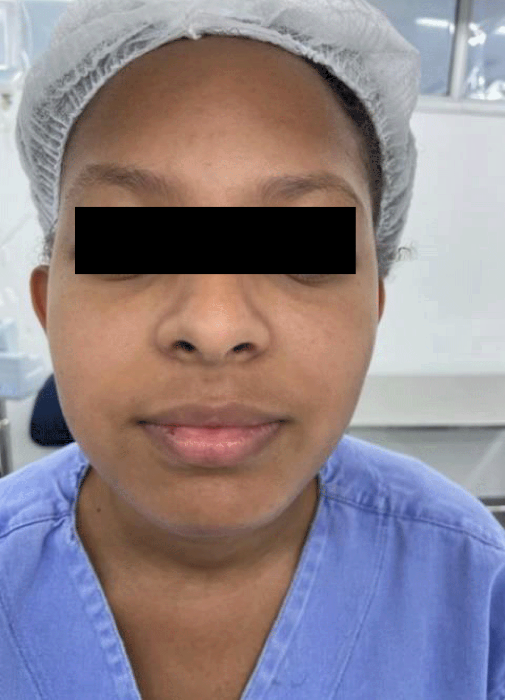
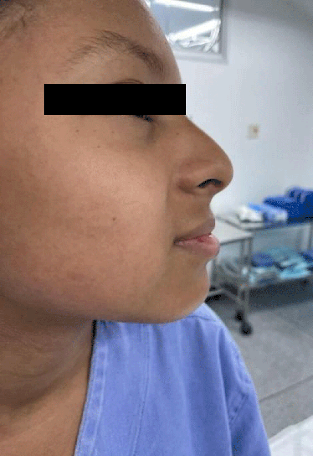
A 15-year-old female patient presented to Darcy Vargas Children’s Hospital with a chief complaint of swelling in the oral mucosa of the anterior mandible. Intraoral examination confirmed dental vitality in the affected area, and the parents reported no history of trauma. Extraoral examination revealed no facial asymmetry (Figures 1,2).
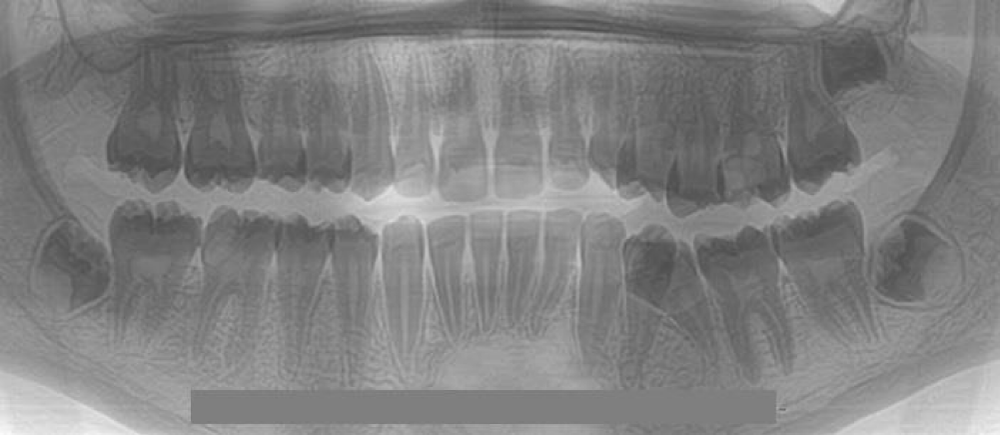
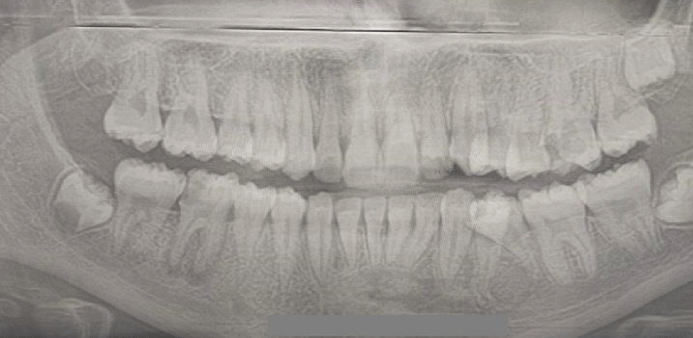
Panoramic X-ray imaging showed a unilocular endosseous lesion located in the chin region between dental elements 22 and 27 (Figures 3,4). The treatment plan included surgical removal of the cyst, curettage of the cystic cavity, and the application of a third-generation PRF membrane to enhance and accelerate osseous healing.
The patient underwent general anesthesia and received antibiotic prophylaxis with Cefazolin at a dose of 30 mg/kg. Postoperative pain management included Ibuprofen 10 ml every 6 hours for five consecutive days.
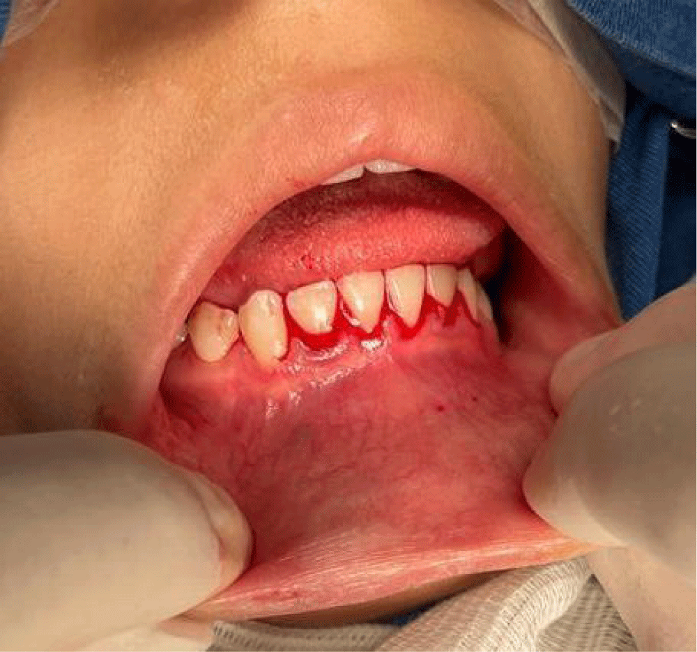
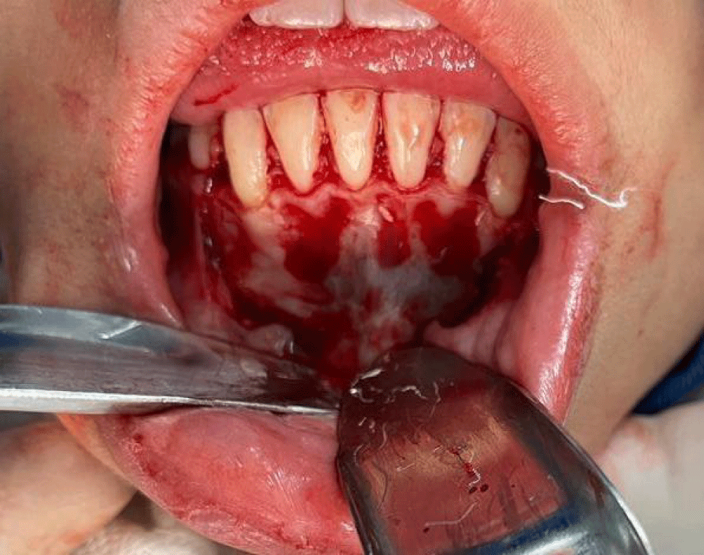
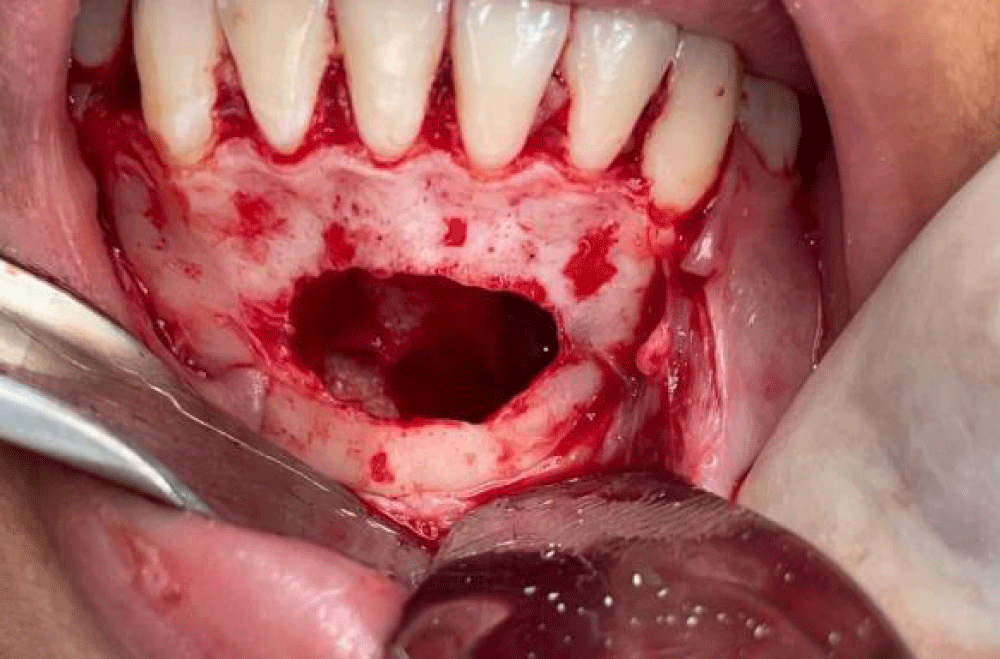
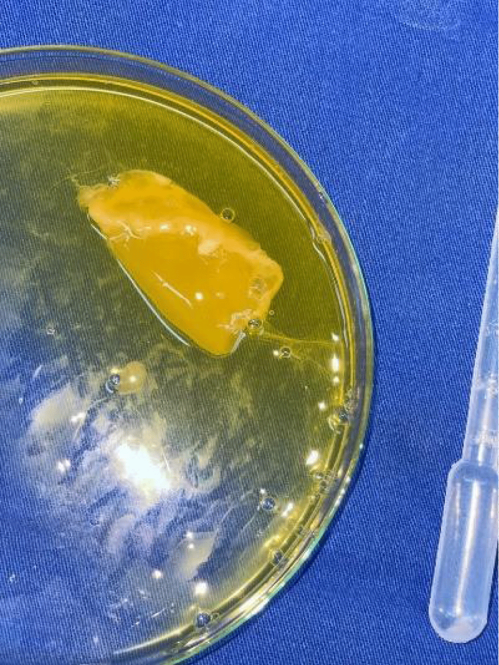
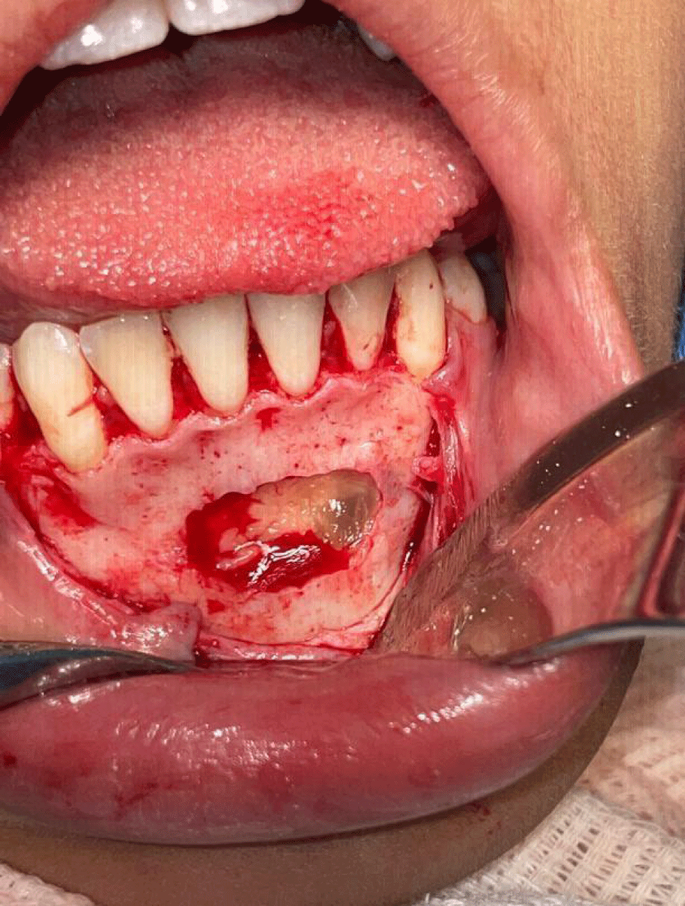
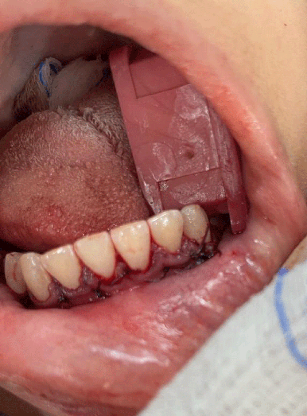
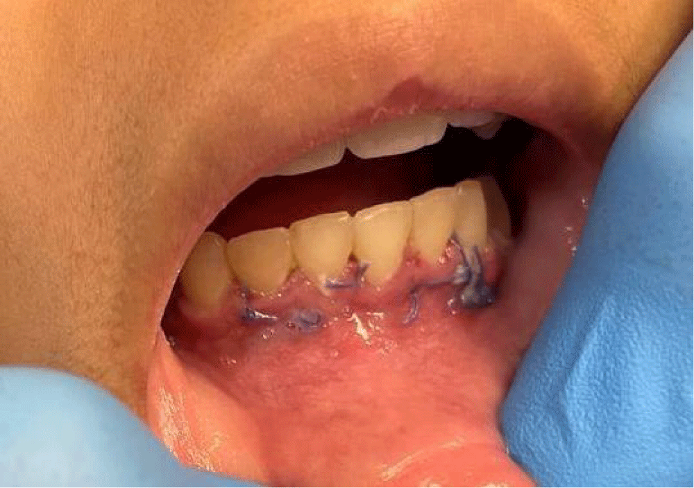
The surgical procedure involved a sulcular incision from the distal papilla of tooth 22 to the distal papilla of tooth 27 (Figure 5), followed by the detachment of mucoperiosteal tissue to expose the lesion and surrounding area (Figure 6). The osteotomy was performed using surgical bur no. 702, and the cystic lesion was removed with a Molt no. 9 periosteal elevator (Figure 7). A PRF membrane was prepared by venipuncture of the patient’s blood, which was collected in vacutainer tubes and centrifuged at 60-700 g for 15 minutes [4]. The resulting blood concentrate was placed in a round glass container, where it polymerized into a membrane after approximately 20 minutes (Figure 8) [5]. This membrane was then inserted into the osseous defect, and the remaining liquid was injected into the area with a plastic pipette to enhance wound healing (Figure 9). The tissue was repositioned, and simple single sutures were placed using monofilament resorbable thread in the papillary area of teeth 22 to 27 (Figure 10).
A follow-up session was conducted for 15 days postoperatively, with no complications observed (Figure 11). A new panoramic X-ray taken six months later showed complete bone healing and new bone formation in the treated area (Figure 12).
In recent times, the domain of wound healing management has evolved, incorporating methodologies centered on tissue engineering. Presently, advancements in biomaterials and a more profound comprehension of the wound-healing cascade have led to the development of numerous innovative therapies and strategies. Regrettably, the majority of these interventions are prohibitively expensive for widespread application across all patient populations and do not consistently ensure efficacy [4].
The present case utilizes an economical adjuvant therapy involving Platelet-Rich Fibrin (PRF), which has demonstrated cost-effectiveness and high efficacy in accelerating the healing process and safeguarding the surgical site against potential postoperative infections [5].
Conclusion
The application of the progressive protocol technique utilizing Platelet-rich Fibrin (PRF) as an accelerator in the wound healing process has demonstrated significant efficacy. This approach not only enhances the speed of healing but also improves the quality of tissue regeneration. The use of PRF in clinical settings, particularly in pediatric patients, has shown promising results in reducing recovery time and promoting optimal healing outcomes. The findings from this case report suggest that PRF can be a valuable adjunct in surgical procedures, offering a reliable method to expedite the healing process and improve patient care. Further studies and clinical trials are recommended to validate these results and explore the full potential of PRF in various dental and medical applications.
Ethical Considerations and Consent form: All signed by the legal responsible (mother) of the patient.
- Saboia-Dantas CJ, Dechichi P, Fech RL, de Carvalho Furst RV, Raimundo RD, Correa JA. Progressive Platelet Rich Fibrin tissue regeneration matrix: Description of a novel, low cost and effective method for the treatment of chronic diabetic ulcers-Pilot study. PLoS One. 2023;18(5):e0284701. Available from: https://doi.org/10.1371/journal.pone.0284701
- Nurden AT. Platelets, inflammation and tissue regeneration. 2011:S13-33. Available from: https://doi.org/10.1160/ths10-11-0720
- Leoni G, NeumannPA, Sumagin R, Denning TL, Nusrat A. Wound repair: role of immune-epithelial interactions. Mucosal Immunol. 2015;8:959-968. Available from: https://doi.org/10.1038/mi.2015.63
- Choukroun J, Ghanaati G. Reduction of relative centrifugation force within PRF (Platelet-Rich-Fibrin) concentrates advances patients’ own inflammatory cells and platelets: First introduction of the Low-Speed Centrifugation Concept, Eur J Trauma Emerg Surg. 2018;44(1):87-95. Available from: https://doi.org/10.1007/s00068-017-0767-9
- Yelamali T, Saikrishna D. Role of platelet rich fibrin and platelet rich plasma in wound healing of extracted third molar sockets: a comparative study. J Maxillofac Oral Surg. 2015;14:410-416. Available from: https://doi.org/10.1007/s12663-014-0638-4
Article Alerts
Subscribe to our articles alerts and stay tuned.
 This work is licensed under a Creative Commons Attribution 4.0 International License.


 Save to Mendeley
