International Journal of Vascular Surgery and Medicine
Management of traumatic recurrent arteriovenous fistula of the lower extremity: Open versus endoscopic repair
Nwafor IA*, Agagwuncha N, Etukokwu E and Eze JC
Cite this as
Nwafor IA, Agagwuncha N, Etukokwu E, Eze JC (2024) Management of traumatic recurrent arteriovenous fistula of the lower extremity: Open versus endoscopic repair. Int J Vasc Surg Med 10(1): 004-008. DOI: 10.17352/2455-5452.000046Copyright License
© 2024 Nwafor IA, et al. This is an open-access article distributed under the terms of the Creative Commons Attribution License, which permits unrestricted use, distribution, and reproduction in any medium, provided the original author and source are credited.Background: Traumatic Arteriovenous Fistula (TAVF) and traumatic artery aneurysms are the late complications of vascular injuries. Complications arise from neglected or undetected cases of TAVF, hence the need to promptly diagnose and manage them.
Aims/Objective: this is to highlight the principles and challenges of managing traumatic recurrent AVF of the lower extremities in a low-income setting like ours. We present a 43- year man with traumatic recurrent AVF of the lower extremity, associated with chronic venous disease and ankle ulcer dating 17 years, who underwent successful treatment via open repair, in our facility, through the combined efforts of a multidisciplinary team involving vascular and plastic surgeons as well as a physiotherapist.
Introduction
Arteriovenous Fistula (AVF) is a direct connection between an artery and vein at a point proximal to the capillary. The causes of this abnormality are threefold and include congenital, therapeutic, and traumatic (iatrogenic and accidental). It is related to arteriovenous malformations but the distinguishing characteristic between the two is the presence of a vascular nidus or nest or epicenter that connects the artery and vein (arteriovenous malformations). It can involve consequential or non-consequential vessels. It may involve central or peripheral vessels.
AVF was first described as an entity in medicine by William Hunter in 1752, followed by the first attempt at surgical correction by Breschet in 1837. He tried to eliminate the fistula by ligation of the proximal or feeder artery [1]. Various factors do play a role in the etiology of AVF, the incidence of which is difficult to determine because of the possibility of delay in diagnosis for years [2,3]. The absence of spontaneous regression within two weeks is an indication of surgical or endovascular treatment [4].
It is one of the most fascinating and misdiagnosed complications of vascular injuries, whether military or civilian [5].
Surgery may involve excision or quadruple ligation if the vessels involved are inconsequential. When the affected vessels are consequential, treatment may involve direct primary repair or anatomical reconstruction using autogenous vein graft, synthetic interposition graft, or bypass [6].
AVF causes venous hypertension, arterializations of the vein wall, varicose veins, and venous ulcers including local gigantism or limb overgrowth. Large AVF causes high-output cardiac failure which may manifest as dyspnoea and peripheral leg edema. Aneurysmal degeneration of the involved artery and venous segments (structural changes) from haemodynamic stress may occur. Total peripheral resistance is lowered distal to the fistula. Subacute Bacterial Endocarditis (SBE) at the site of the fistula and venous insufficiency including peripheral ischemia may occur as complications [6-8].
Case summary
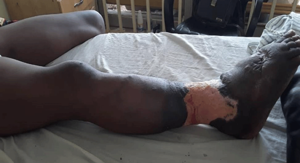
A 43-year-old commercial bus driver presented to our service with a two-month history prior, to a fifteen-year history of recurrent right leg swelling (Figure 1). His problem, when it started was insidious in onset. The swelling initially started in the foot and later deteriorated to involve the leg and the thigh. There were associated varicose veins and circumferential ankle ulcers. There was associated pain, worsened by activities like standing and walking and relieved by analgesics. There was a preceding history of gunshot injury to the right lower limb two months before the onset of symptoms. He initially presented to a peripheral hospital where he was evaluated and diagnosed as a case of traumatic arteriovenous fistula, after digital subtraction angiography. Repair of the AVF was done and the patient had an uneventual recovery. However, three months later, the symptoms relapsed, necessitating the patient’s presentation to our service, for expert management.
Examination revealed a middle-aged man in no obvious distress, afebrile, severely pale, anicteric, acyanosed, not dehydrated, nil peripheral edema of the contralateral limb. The musculoskeletal evaluation showed the right lower limb that was swollen up to the mid-thigh with hyperpigmentation and engorged tortuous veins noted around the upper two-thirds (2/3) of the leg. The oedema was non-pitting. There was a tender circumferential ulcer at the ankle, with a sloping edge and floor oozing seropurulent, foul-smelling discharge. All peripheral arterial pulsations and skin sensations were intact. There was a significant thrill felt over the mid-thigh extending to the femoral triangle. The bruit was positive over the sites of the thrill. However, bruit was more pronounced at the mid-thigh. There was no abnormality in the contralateral limb.
The leg is swollen circumferential ankle ulcer(hypopigmented) and swollen, elephantoid foof
Thigh circumferences (30 cm from the anterior superior iliac spine): (R) is 59 cm while (L) is 54.5cm. Leg circumferences (15cm from tibial tuberosity): (R) is 42.5 cm while (L) is 38 cm.
In the cardiovascular system examination pulse rate was found to be 89 pulsations per minute, full volume, and regular rhythm. All the peripheral pulses were present and synchronous. Blood pressure was 110/60 mmHg. There was no raised jugular venous pressure. The apex beat was on the 5th left intercostal space, midclavicular line. Heart sounds, S1&S2 were heard, and no murmur.
Respiratory system examination showed that respiratory rate was 20 cycles per minute and SPO2 on room air was 98%. Auscultations revealed a vesicular breath sound and no added sound.
There were no significant findings in the abdominal and urogenital examinations respectively.
Confirmatory Investigations (Doppler ultrasound) were not helpful as it demonstrated as follows: Artery and vein in the distal third of the thigh with suspected right arteriovenous fistula involving the superficial femoral artery with areas of AV fistula in the pelvic cavity (external iliac artery and vein and common femoral artery and vein on the right). There were prominent superficial veins in the right which suggest an inflammatory response rather than venous insufficiency.
However, the computerized tomography scan showed the following: Early enhancement of the right external iliac vein, common femoral vein, and superficial femoral vein with focal berry aneurysmal dilatation is demonstrated.
The right common femoral vein, superficial vein, and its corresponding arteries appear engorged relative to the contralateral lower limb with an abrupt change in the caliber noted at the level of proximal 2/3rd slightly above the knee.
Extensive vascular nidus within the soft tissues of the medial right thigh is noted with the arterial supply arising from the small branches of the femoral artery and profunda femoris.
The left lower limb appears to be normal in its vascular configuration.
There is an asymmetry in the size of the lower limb (RT>LT) consistent with right hemihypertrophy.
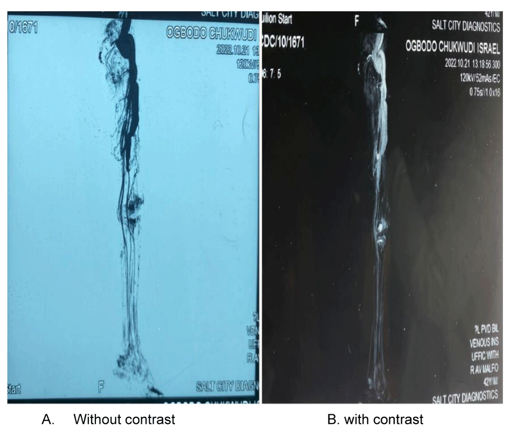
The contrast outlines the upper vasculature femoral artery with abrupt disappearance in the distal third of the femur
Based on the clinical evaluation with the investigations, a working diagnosis of traumatic recurrent right lower limb Arteriovenous Fistula (AVF) complicated with Chronic Venous Disease (CVD) and ankle ulcer was made.
Treatment was multidisciplinary involving vascular and plastic surgeons as well as physiotherapists. The patient was admitted into the ward and treated with parenteral antibiotics and analgesics. He was transfused 2 units of blood (post-transfusion hemoglobin was 11.3g/dl). Wound care was also commenced by the plastic surgery unit. He later had surgical repair of the AV fistula on the 9th of November, 2023.
Intra-operative details
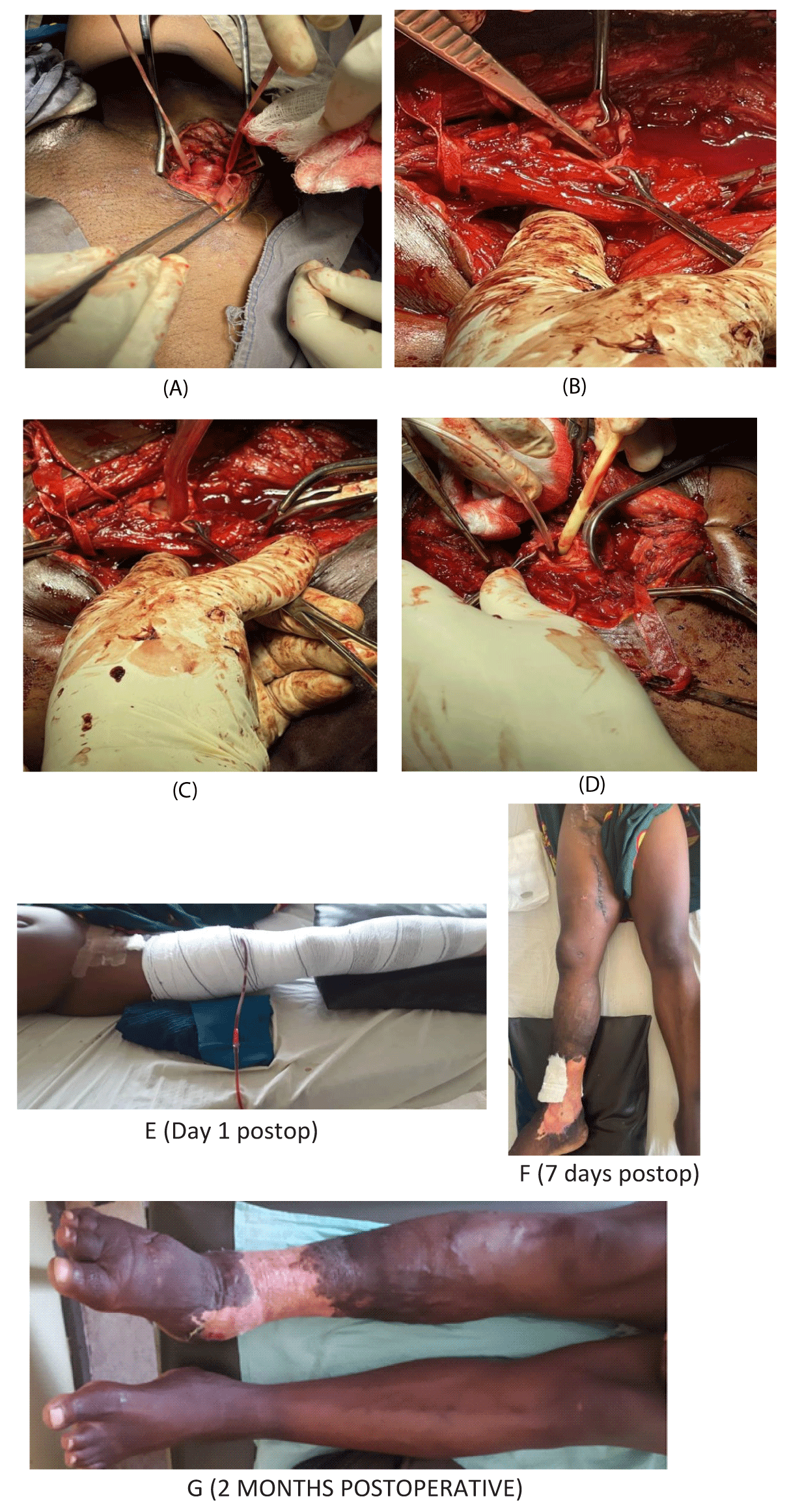
The right lower limb is still swollen but has improved substantially. Circumferential right ankle ulcer showed healthy granulation. Thigh circumferences: (30 cm from Anterior superior iliac spine): Right – 56 cm, Left – 54.5 cm. Leg circumferences: (15 cm from tibial tuberosity) Right – 40 cm, Left – 38 cm.
The specific recovery status included the rapid healing of the ulcer, and regression of the distended, tortuous, and elongated superficial veins (varicose) including a progressive reduction in the segmental diameters of the thigh and leg as illustrated in Table 1.
Discussion
Vascular injuries caused by civilian or war trauma represent a surgical challenge. These lesions are potentially lethal and can cause death at the scene. One of the most fascinating and misdiagnosed complications of vascular injuries is the Arteriovenous Fistula (AVF), which results from direct communication between an artery and a vein.[9] They are usually secondary to penetrating trauma and occasionally may be diagnosed many years after the injury. AVF represents a significant diagnostic and management challenge. Angiography is necessary to identify the affected vessels as this helps to develop a surgical treatment plan (open/close/combination) [10]. Comprehensive clinical cum investigative review helps to do hemodynamic analysis and enables the choice of either an open or closed approach. In this index case preoperative computerized angiography, both contrast-enhanced and non-contrast-enhanced provides the site of AVF around the distal 3rd of the femoral artery. Figures 2A,B.
When AVF presents with complications, multidisciplinary approaches are needed and they may involve vascular and plastic surgeons, hematologist cardiologists, interventional radiologists, and physiotherapists. In the index case, vascular and plastic surgeons as well as physiotherapists were involved in the management.
In the index case, the specific operation was to get proximal control at the level of the femoral artery and distal control at the level of the anterior and posterior tibia arteries. Figure 3C. After that, an arteriotomy was done and the site of the fistula was identified, a size 16 FG Foley catheter was passed through the fistula and inflated to occlude and achieve hemostasis. Similarly, a size 16 nasogastric tube was passed through the distal lumen. Figure 3D. The fistula was then closed with prolene 3/0 which caused the disappearance of the thrill when the proximal clamp was released for testing. Subsequently, the arteriotomy was closed and later, the wound was closed in layers with an active drain in situ.
The objectives borne in mind in the management are to close the AVF, maintain the patency of the consequential vessels, and exclude or excise the non-consequential vessels as well as decrease or eliminate several complications such as cardiac failure, venous hypertension, and local gigantism among others [11]. In the index case, there was evidence of a reduction in local gigantism after repair. Table 1.
The principles of treatment are sequential and include obtaining proximal and distal control, wide exposure over the point where the thrill is palpated highest or the area of the AVF, as outlined by preoperative angiography, Systemic heparinization, and arteriotomy for closure of the AVF under the clear vision and then the arteriotomy [12]. In the index patient, the femoral artery and vein are consequential vessels; the above principles were followed to the later. Figures 3A-D. Unlike the index case, where the communication site is complex, the artery should be ligated and reconstruction effected using autogenous vein graft or prosthesis. Bypass can also be done [13].
The open approach may be fraught with danger owing to grossly distorted and edematous tissue planes and late presentation of AVF is prone to significant intra-operative bleeding due to the complex vascular anatomy encountered during surgical dissection and repair [14]. In the index case, there was dense adhesion and venous hypertension that led to significant intraoperative blood loss. To avoid morbidity and mortality associated with an open procedure, emphasis is now on the endovascular approach.
In developed centers unlike ours, with equipment and expertise to manage this type of AVF, the objectives are usually to selectively eliminate the AVF via an endoscopic approach with preservation of normal patency of consequential vessels (femoral artery and vein) and reduce morbidity and mortality encountered in open repair. In addition to this inherent advantage, there is rapid postoperative recovery and a reduction in pain and disability. Available options in endoscopic treatment are the use of covered stents (expandable or non-expandable), coil embolization, and balloon occlusion techniques. It is noteworthy that biodegradable and retrievable stents are now available for use in cutting-edge technology to manage challenging AVFs, especially in children [15,16].
The advantages of applications of endoscopic repair have inherent advantages and they include reduced pain postoperatively, reduced morbidity, and rapid recovery. The disadvantages are occasional rupture of a vessel or dissection and embolization of the devices leading to distal ischemia.
On the other hand, the following factors will make open treatment inevitable: Discrepancy between proximal and distal diameters of the vessel, the impossibility of catheterization of the vessel, Injuries that need exploration, hematoma with compressive symptoms and infected wounds [17,18]. The index case was suited for endovascular treatment, but our center lacked the technical know-how.
Conclusion
Management of large AVF of the lower extremity, with prolonged duration, where consequential vessels are involved, not to mention recurrent ones, complicated with chronic venous disease and ulcer, is very challenging. However, the adoption of standard principles coupled with the expertise of a competent vascular surgeon alongside other multidisciplinary teams, there was the resolution of symptoms with the patient recovering satisfactorily. The absence of an endoscopic treatment facility in our facility sometimes creates avoidable morbidity like severe postoperative pain, delayed recovery, and prolonged hospital stay.
- Grace C, Andrew BQ, Brasmo SDS. Traumatic Arteriovenous Fistula. Inetech Open chapter 10. http://dx.doi.org/10.5772/56368.
- Stathis A, Gan J. Traumatic arteriovenous fistula: a 25-year delay in presentation. J Surg Case Rep. 2020 Mar 24;2020(3):rjaa042. doi: 10.1093/jscr/rjaa042. PMID: 32226601; PMCID: PMC7092680.
- Nagpal K, Ahmed K, Cuschieri R. Diagnosis and management of acute traumatic arteriovenous fistula. Int J Angiol. 2008 Winter;17(4):214-6. doi: 10.1055/s-0031-1278313. PMID: 22477453; PMCID: PMC2728918.
- Spencer TA, Smyth SH, Wittich G, Hunter GC. Delayed presentation of traumatic aortocaval fistula: a report of two cases and a review of the associated compensatory hemodynamic and structural changes. J Vasc Surg. 2006 Apr;43(4):836-40. doi: 10.1016/j.jvs.2005.12.003. PMID: 16616246.
- Huang W, Villavicencio JL, Rich NM. Delayed treatment and late complications of a traumatic arteriovenous fistula. J Vasc Surg. 2005 Apr;41(4):715-7. doi: 10.1016/j.jvs.2005.01.049. PMID: 15874939.
- Martinez L, Li agostera S, E Sandera JR. Open Surgical Repair of an iatrogenic popliteal arteriovenous fistula with proximal arteriomegaly: A case report. Ejves Extra(Elsevier) 2011;22(^):e70-72.
- Khoury G, Sfeir R, Nabbout G, Jabbour-Khoury S, Fahl M. Traumatic arteriovenous fistulae: "the Lebanese war experience". Eur J Vasc Surg. 1994 Mar;8(2):171-3. doi: 10.1016/s0950-821x(05)80454-2. PMID: 8181610.
- Fox CJ, Gillespie DL, O'Donnell SD, Rasmussen TE, Goff JM, Johnson CA, Galgon RE, Sarac TP, Rich NM. Contemporary management of wartime vascular trauma. J Vasc Surg. 2005 Apr;41(4):638-44. doi: 10.1016/j.jvs.2005.01.010. PMID: 15874928.
- Kelm M, Perings SM, Jax T, Lauer T, Schoebel FC, Heintzen MP, Perings C, Strauer BE. Incidence and clinical outcome of iatrogenic femoral arteriovenous fistulas: implications for risk stratification and treatment. J Am Coll Cardiol. 2002 Jul 17;40(2):291-7. doi: 10.1016/s0735-1097(02)01966-6. PMID: 12106934.
- HUGHES CW. Arterial repair during the Korean war. Ann Surg. 1958 Apr;147(4):555-61. PMID: 13521671; PMCID: PMC1450643.
- Carvajal G, Brito A, Simo E. Traumatic arteriovenous fistula: Diagnosis and Management, Intech open 2013; http://dx.doi.org/org/10.5772/56368.
- Ezemba N, Ekpe EE, Ezike HA, Anyanwu CH. Traumatic common carotid-jugular fistula: report of 2 cases. Tex Heart Inst J. 2006;33(1):81-3. PMID: 16572879; PMCID: PMC1413591.
- Kaklikkaya I, Ozcan F, Kutlu N. Double layered autogenous vein graft patch reconstruction of the common carotid-internal jugular fistula caused by gunshot wound. J Cardiovasc Surg (Torino). 1999 Jun;40(3):429-33. PMID: 10412935.
- Anyanwu CH, Ude AC, Swarup AS, Umerah BC, Udekwu FA. Traumatic aneurysms and arteriovenous fistulas in Nigeria. Thorac Cardiovasc Surg. 1980 Aug;28(4):265-8. doi: 10.1055/s-2007-1022092. PMID: 6158130.
- Adeyemi-Duro HO. Traumatic aneurysms and arteriovenous fistula of the upper limb. West Afr J Med. 1989 Apr-Jun;8(2):143-9. PMID: 2486787.
- Anderson CA, Strumpf RK, Diethrich EB. Endovascular management of a large post-traumatic iliac arteriovenous fistula: utilization of a septal occlusion device. J Vasc Surg. 2008 Dec;48(6):1597-9. doi: 10.1016/j.jvs.2008.06.065. PMID: 19118742.
- Topuz M, Coşgun M, Şen Ö, Çaylı M. Coil embolization of a traumatic arteriovenous fistula of the lower extremity. Turk Kardiyol Dern Ars. 2015 Dec;43(8):724-6. doi: 10.5543/tkda.2015.44045. PMID: 26717336.
- Carvalho Cla, Silva Ln, Lima Mbc, Cavalcante Rcr, Vasconcelos-Filho Jom. Traumatic arteriovenous fistula: a literature review. Clin Surg J. 2021;2(3):1-10.
Article Alerts
Subscribe to our articles alerts and stay tuned.
 This work is licensed under a Creative Commons Attribution 4.0 International License.


 Save to Mendeley
